Knee Meniscus Injuries
Posted on February 12, 2023 by Movement Health in Exercise Physiology, Just saying..
What is the meniscus?
Meniscus are crescent shaped fibrocartilaginous structures that are located between the femur (thigh bone) and tibia (shin bone), they are located within the knee joint (tibiofemoral joint) and are attached to the joint capsule at the knee joint line. Each knee has two menisci, towards the inner aspect of the knee is the medial meniscus and towards the outer aspect of the knee is the lateral meniscus. Meniscus primarily function as a knee joint shock absorber between the articular surfaces of the femur and tibia, meniscus also function as a means of transmitting force through the knee joint, stabilising the knee joint and lubricating the knee joint (Cooper et al. 2009).
Types of knee meniscus injuries
Meniscal injuries are common knee injuries and can be classified as acute or degenerative.
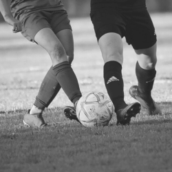
Acute meniscal injuries are typically seen in young athletes as a result of a large shear load being put through an otherwise healthy knee meniscus (Thorland et al. 2018). The typical mechanism for an acute meniscal injury is a knee that is flexed and in compression experiencing rotational forces (Cooper et al. 2009).
Degenerative meniscal injuries are more common in middle-aged and older individuals and can often be present alongside knee osteoarthritis. Degenerative meniscal injuries often don’t have a mechanism of injury (Thorland et al. 2018) and may be considered a part of, ‘the timeline of life’.
Signs and Symptoms
Acute meniscal injuries will often present as a knee that is painful when bending, straightening, or twisting, alongside a generally restricted knee range-of-motion. Knee joint effusion or accumulation of fluid around the knee joint can be another sign of a meniscal injury. Small (acute) meniscal tears can demonstrate delayed effusion, this is joint effusion that only becomes obvious 6-24 hours post injury. More severe (acute) meniscal injuries may also present as a knee that makes a ‘clicking’ or ‘popping’ noise, more severe (acute) meniscal injuries may also ‘lock’ at various joint angles (Cooper et al. 2009).
Degenerative meniscal injury symptoms may present gradually as a knee that is painful and weak, the weakness will often be experienced with walking endurance and balance. The knee experiencing a degenerative meniscal injury will typically demonstrate a range-of-motion that is equal to the contralateral knee and typically there is nil presence of knee joint effusion. People experiencing degenerative meniscal injury will often report a knee that ‘clicks’, ‘pops’, ‘locks’ or ‘gives way’ (Howell et al. 2014).
What to expect from your healthcare provider?
If you’re experiencing a knee meniscus injury, you’ll likely need to work with someone who has a good understanding of health science, pain and exercise, this could include health professionals such as a Sports Physician, Exercise Physiologist (learn more here), Physiotherapist or Chiropractor. Most importantly you need to feel like you have a good rapport with your chosen healthcare provider, so it’s ok to ask around and pick the professional that is the right fit for you.
Acute meniscal injuries
For an acute meniscal injury your health professional will likely start by asking you to share your experiences and any information about the injury, things they may be interested in could be:
- A description of how the knee injury happened.
- Is the knee painful when bending/straightening/twisting?
- Does the knee click, pop or lock?
A common description from people who have acutely injured their knee meniscus whilst participating in sport is an experience of twisting the knee while the foot is planted (Cooper et al. 2009).
Next your health professional will take an objective look at the knee, during this process they will look at all the structures of the knee. Other knee structures they may be interested in examining could be the ACL and MCL. The objective knee assessment will likely include examination of the knees range-of-motion, pain levels, the presence of effusion/swelling, and the ability of the knee to bear load and functionally bend (squat) (Cooper et al. 2009). Orthopaedic tests that are commonly used to examine the knee meniscus are Joint Line Tenderness and the McMurrays test; other less utilised tests include the Apley’s and Thessaly’s tests (Smith et al. 2015). Orthopaedic tests cannot give a thorough picture around the integrity of a meniscal injury like an MRI can, however your health professional will look at a ‘basket’ of presentations to establish a knee meniscus injury diagnosis, this ‘basket’ could include:
- Twisting mechanism of injury.
- Knee joint effusion or accumulation of fluid around the knee joint.
- Delayed knee joint effusion.
- Reduced knee range-of-motion.
- Knee pain with bending/squatting.
- Knee that clicks, pops or locks.
- Knee joint line tenderness.
- Positive McMurray’s test (Cooper et al. 2009).
Degenerative meniscal injuries
For a degenerative meniscal injury, your health professional will likely start by asking you to share your experiences and any information about the knee, things they may be interested in could be:
- A description around your experiences with the knee.
- Does the knee feel weak or painful?
- How does the knee handle walking long distances?
- What are some tasks that may irritate the knee?
- Does the knee stop you participating in tasks that are meaningful? What are those tasks?
- Is the knee painful when bending/straightening/twisting?
- Does the knee click, pop, lock or give way?
Next your health professional will undertake an objective assessment of the knee, examining all its structures so that any other pathologies can be excluded. Joint effusion is rarely present in knees experiencing a degenerative meniscal injury and often the injured knees range-of-motion will be equal to the uninjured knee. The Joint Line Tenderness and McMurray’s orthopaedic test may also be used to develop a picture around the integrity of the degenerative meniscus. The degenerative meniscal injuries diagnostic ‘basket’ may include:
- Person experiencing knee pain is middle-aged or older.
- Gradual onset of knee pain.
- Nil recollection of a mechanism that may have injured the knee.
- Presence of knee osteoarthritis.
- Knee weakness compared to the uninjured knee.
- Decreased walking endurance.
- Decreased balance.
- Knee that clicks, pops, locks, or gives way.
- Knee joint line tenderness.
- Positive McMurray’s test (Howell et al. 2014)
Treatment – Acute meniscal injuries
Initial acute meniscal injury management should include the ‘PEACE and LOVE’ protocol (learn more here) for soft tissue injury (Dubois & Esculier, 2020). Once the knee is ‘quiet’ treatment for acute meniscal injuries can vary between conservative and surgical interventions. Meniscal surgery is typically performed via small incisions on the anterior (front) surface of the knee through which a small camera called an arthroscope and various surgical tools are passed (Cooper et al. 2009). Typical meniscal surgeries may include a partial meniscectomy, where the loose piece of meniscus is removed or a meniscal repair where the loose piece of meniscus is sutured back into place (Smoak et al. 2020). Conservative interventions may involve exercises focused on restoration of knee range-of-motion, muscle function and coordination. Exercise programming should be graded towards daily tasks or athletic performance (Cooper et al. 2009) and functional performance tests that are commonly used to monitor progress are the single leg hop and the crossover hop tests (Berg et al. 2022).
There is limited evidence around what is the best treatment approach for managing acute meniscal injuries (Culvenor et al. 2022; Thorland et al. 2018), therefore it is important to work with health professionals who can help to negotiate the ‘grey zones’. A good approach may be to start with conservative management, delay any discussions about surgery and if the knee doesn’t respond undertake surgery at a later date (Thorlund et al. 2018; van Der Graff et al. 2022). Some considerations when exploring rehabilitation options may include:
- Acute meniscal damage can take various forms, tears can be a flap, radial, longitudinal, bucket handle or cleavage (Cooper et al. 2009). Longitudinal tears to the outer parts of the meniscus are good candidates for a suture repair, as this part of the meniscus is more vascular and therefore better inclined towards healing (Smoak et al. 2020). Inner parts of the meniscus have limited blood supply and typically a meniscectomy will be undertaken (Cooper et al. 2009).
- The typical indications for meniscal surgery are an MRI that demonstrates damaged meniscal tissue and the presence of mechanical symptoms (Thorlund, 2018). Mechanical symptoms are when an injured knee ‘catches’ or ‘locks’, however the relationship between these symptoms and a positive MRI is not well understood (Thorlund, 2022), therefore it is advised to collaborate with your chosen health professionals while you negotiate the ‘grey zone’.
- It is generally accepted that younger people will respond better to meniscal surgery following acute damage, however this relationship is not well understood (Thorlund, 2018), therefore it’s advised to collaborate with your chosen health professionals regarding any decision making about age.
Treatment – Degenerative meniscal injuries
Arthroscopic partial meniscectomy surgery is typically performed to manage degenerative meniscal injuries, however there is limited evidence supporting the effectiveness of this procedure (Migliorini et al. 2022; Thorlund et al. 2015). Exercise therapy has been shown to be equal to arthroscopic surgery in reducing knee pain and improving knee function; and exercise therapy is more effective at developing knee strength (Thorlund et al. 2015). Therefore, best practice management for degenerative meniscal knee injuries should be non-surgical and prioritise exercise therapy (Noorduyn et al. 2022; Thorlund et al. 2018).
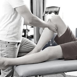
So what might exercise therapy look like in practice?
Initially it is best to seek out a health professional with whom you share a good rapport, this will support an environment where you can collaborate on a plan for improved knee health. Exercise programming can take many forms and it’s important your health professional listens to your experiences and helps develop a strategy that aligns with these experiences. It’s important you have input into your exercise programming and feel like you are working towards meaningful goals. Your health professional should be able to explain what a meniscal (acute or degenerative) injury is, in language you understand, knowledge is power and understanding your health is a part of the process when improving your health. You should feel safe to ask questions about your rehabilitation, your exercise programming should be graded/progressed over time, and you should be developing skills that allow you to eventually self-manage your exercise programming.
Points to consider
- Acute meniscal injuries have nil threshold for surgery, the decision to operate typically relates to professional opinion.
- There’s nil advantage for knee health to be achieved via arthroscope surgery for a degenerative meniscal injury. Exercise/physical activity should be prioritised.
- Seek out an exercise-informed health professional whom you can collaborate with across the rehabilitation timeline.
Thanks for reading, Warwick..
(If you found this article helpful and would like to support my writing, you could, shout me a coffee).
Berg, B., Urhausen, A.P., Øiestad, B.E., Whittaker, J.L., Culvenor, A.G., Roos, E.M., Crossley, K.M., Juhl, C.B., & Risberg, M.A. (2022) What tests should be used to assess functional performance in youth and young adults following anterior cruciate ligament or meniscal injury? A systematic review of measurement properties for the OPTIKNEE consensus. British Journal of Sports Medicine, 56(24), 1454-1464.
Cooper, R., Morris, H., & Arendt, L. (2009). Acute Knee injuries. In P. Brunker & K. Kahn (Eds.), Clinical Sports Medicine Revised Third Edition (pp. 468-472). Mc Graw Hill.
Culvenor, A.G., Girdwood, M.A., Juhl, C.B., Patterson, B.E., Haberfield, M.J., Holm, P.M., Bricca, A., Whittaker, J.L., Roos, E.M., & Crossley, K.M. (2022). Rehabilitation after anterior cruciate ligament and meniscal injuries: a best-evidence synthesis of systematic reviews for the OPTIKNEE consensus. British Journal of Sports Medicine, 56(24), 1445-1453.
Dubois, B., & Esculier, J. (2020). Soft-tissue injuries simply need PEACE and LOVE. British Journal of Sports Medicine, 54(2), 72-73.
Howell, R., Kumar, N.S., Patel, N., & Tom, J. (2014). Degenerative meniscus: Pathogenesis, diagnosis, and treatment options. World Journal Orthopedics 5(5), 597–602.
Migliorini, F., Oliva, F., Eschweiler, J., Cuozzo, F., Hildebrand, F., & Maffulli, N. (2022). No evidence in support of arthroscopic partial meniscectomy in adults with degenerative and nonobstructive meniscal symptoms: a level I evidence-based systematic review. Knee Surgery, Sports Traumatology, Arthroscopy. Epub ahead of print. doi: 10.1007/s00167-022-07040-0
Noorduyn, J.C.A., van de Graaf, V.A., Willigenburg, N.W., Scholten-Peeters, G.G.M., Kret, E.J., van Dijk, R.A., Buchbinder, R., Hawker. G.A., Coppieters, M.W., & Poolman, R.W. (2022). ESCAPE Research Group. Effect of Physical Therapy vs Arthroscopic Partial Meniscectomy in People With Degenerative Meniscal Tears: Five-Year Follow-up of the ESCAPE Randomized Clinical Trial. Journal of the American Medical Association Network Open, 5(7), e2220394.
Smith, B.E., Thacker, D., Crewesmith, A., and Hall, M. (2015). Special tests for assessing meniscal tears within the knee: a systematic review and meta-analysis. Evidence Based Medicine 20(3), 88-97.
Smoak, J.B., Matthews, J.R., Vinod, A.V., Kluczynski, M.A., & Bisson, L.J. (2020). An Up-to-Date Review of the Meniscus Literature: A Systematic Summary of Systematic Reviews and Meta-analyses. Orthopaedic Journal of Sports Medicine, 8(9), 1-14.
Thorlund, J.B., Juhl C.B., Ingelsrud, L.H., & Skou, S.T. (2018). Risk factors, diagnosis and non-surgical treatment for meniscal tears: evidence and recommendations: a statement paper commissioned by the Danish Society of Sports Physical Therapy (DSSF). British Journal of Sports Medicine, 52(9), 557-565.
Thorlund, J.B. (2022). Meniscal Tears And ‘Mechanical Symptoms’ – An Unsolved Puzzle. Retrieved February 12, 2023 from: https://www.physio-network.com/blog/meniscal-tears-and-mechanical-symptoms-an-unsolved-puzzle/
Thorlund, J.B., Juhl, C.B., Roos, E.M., & Lohmander, L.S. (2015). Arthroscopic surgery for degenerative knee: systematic review and meta-analysis of benefits and harms. British Journal of Sports Medicine, 49(19), 1229-1235.
van der Graaff, S.J.A., Eijgenraam, S.M., Meuffels, D.E., van Es, E.M., Verhaar, J.A.N., Hofstee, D.J., Auw Yang, K.G., Noorduyn, J.C.A., van Arkel, E.R.A., van den Brand, I.C.J.B., Janssen, R.P.A., Liu, W.Y., Bierma-Zeinstra, S.M.A., & Reijman, M. (2022). Arthroscopic partial meniscectomy versus physical therapy for traumatic meniscal tears in a young study population: a randomised controlled trial. British Journal of Sports Medicine, 56(15), 870–876.